Home
Uncategories
Anatomy Of Chest : Bodybuilding chest exercise and anatomy - YouTube - It is enclosed by the ribs, the vertebral column, and the sternum, or breastbone, and is separated from the abdominal cavity (the body's largest hollow space) by a muscular and membranous partition, the diaphragm.
Anatomy Of Chest : Bodybuilding chest exercise and anatomy - YouTube - It is enclosed by the ribs, the vertebral column, and the sternum, or breastbone, and is separated from the abdominal cavity (the body's largest hollow space) by a muscular and membranous partition, the diaphragm.
Anatomy Of Chest : Bodybuilding chest exercise and anatomy - YouTube - It is enclosed by the ribs, the vertebral column, and the sternum, or breastbone, and is separated from the abdominal cavity (the body's largest hollow space) by a muscular and membranous partition, the diaphragm.. Your sternum protects the organs of your torso from injury and also serves as a. The chest or thorax region of the upper body has a number of important organs that reside within it that may present with chest pain if they become compromised in. The superior thoracic aperture found superiorly and the inferior thoracic aperture. Radiology basics of chest ct anatomy with annotated coronal images and scrollable axial images to help medical students and junior doctors learning anatomy. It is enclosed by the ribs, the vertebral column, and the sternum, or breastbone, and is separated from the abdominal cavity (the body's largest hollow space) by a muscular and membranous partition, the diaphragm.
The human thorax includes the thoracic cavity and the thoracic wall. Get the latest information on lungs function now. It provides protection to vital organs (eg, heart and major vessels, lungs, liver) and provides stability for movement. Swensen fund for innovation in teaching. The chest is made up primarily of two muscles:
How To Paddle Stronger and Longer from surfstrengthcoach.com This chapter is an abbreviated review of thoracic anatomy as seen on chest radiographs and computed tomography (ct) of the chest. This atlas is a comprehensive and affordable learning tool for medical students and residents and especially for radiologists and pneumologists. Your sternum protects the organs of your torso from injury and also serves as a. It's also sometimes referred to as the breastbone. The shape of the chest is often regarded as potential insight into a disease process, as in the case of barrel chest and respiratory dysfunction. Learn about each of these muscles, their locations, functional anatomy and exercises for them. Swensen fund for innovation in teaching. Related posts of anatomy of the chest area pressure points on female anatomy.
Get the latest information on lungs function now.
A new and simple method will help you to lose weight fast. 12 cm (5 in) in length, 8 cm (3.5 in) wide, and 6 cm (2.5 in) in thickness. Anatomy of right side chest pain. Follow the steps to lose weight fast. Pressure points on female anatomy 11 photos of the pressure points on female anatomy female dog names, female pleasure points, female pressure points diagram, male pressure points, pressure points on female body, human anatomy, female dog names, female pleasure points, female pressure points diagram, male. Applied anatomy of the chest wall and mediastinum petros mirilas michael e. Chest a man's chest — like the rest of his body — is covered with skin that has two layers. Thoracic cavity, also called chest cavity, the second largest hollow space of the body. A typical heart is approximately the size of your fist: It's also sometimes referred to as the breastbone. Here, we break down the anatomy of your chest muscles. The thoracic skeleton creates a protected space for the heart. Related posts of anatomy of the chest area pressure points on female anatomy.
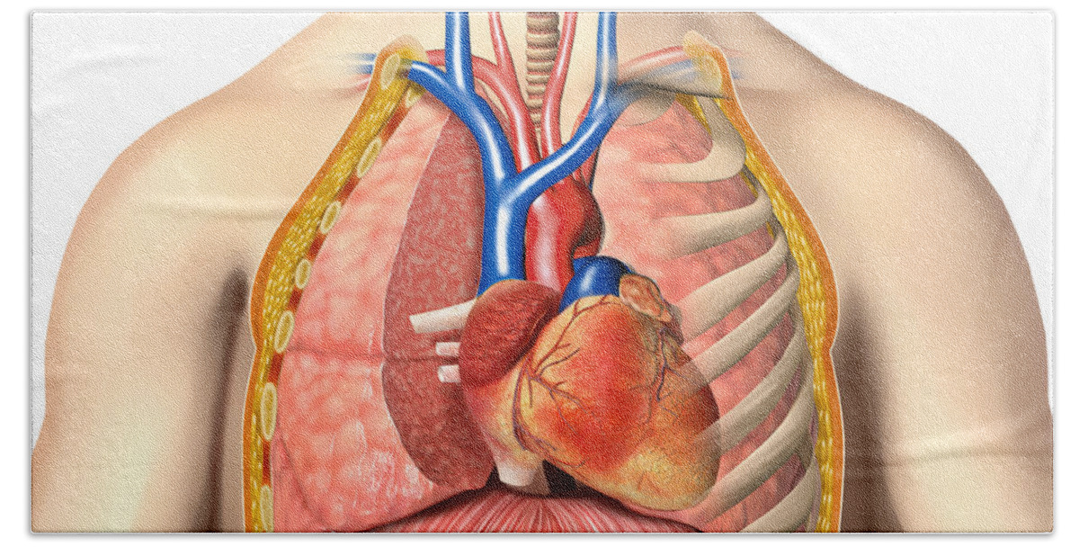
Having to do with the chest. Your sternum is a bone that's located in the middle of your chest. It provides access to ct images in the axial plane, allowing the user to learn and review the lung anatomy interactively. Anatomically, the heart is located in the anterior thoracic cavity; The chest wall is comprised of skin, fat, muscles, and the thoracic skeleton.
Male Chest Anatomy Of Thorax Bath Towel for Sale by Leonello Calvetti from render.fineartamerica.com This atlas is a comprehensive and affordable learning tool for medical students and residents and especially for radiologists and pneumologists. A new and simple method will help you to lose weight fast. A good radiologist knows the anatomy because knowing where structures normally live and recognizing the location of an abnormality helps to make or narrow the differential diagnosis. Definition (nci_cdisc) the anterior side of the thorax from the neck to the abdomen. Your sternum is a bone that's located in the middle of your chest. In insects, crustaceans, and the extinct trilobites, the thorax is one of the three main divisions of the creature's body, each of which is in turn composed of multiple segments. Chest muscles anatomy (1) pectoralis major muscle. Anatomy of the thorax shari l.
Pressure points on female anatomy 11 photos of the pressure points on female anatomy female dog names, female pleasure points, female pressure points diagram, male pressure points, pressure points on female body, human anatomy, female dog names, female pleasure points, female pressure points diagram, male.
How to view the anatomical labels. This chapter is an abbreviated review of thoracic anatomy as seen on chest radiographs and computed tomography (ct) of the chest. Structures to identify • heart • lungs • mediastinum • pleural space • chest wall • …everything else! The chest anatomy includes the pectoralis major, pectoralis minor and the serratus anterior. Learn about each of these muscles, their locations, functional anatomy and exercises for them. The pec major) is the one that commands the most real estate. The chest or thorax region of the upper body has a number of important organs that reside within it that may present with chest pain if they become compromised in. A new and simple method will help you to lose weight fast. Studied the anatomy of the breast, its topography, innervation, vascularization and lymphatic drainage, and correlated the anatomical data with the classification of lymph node groups that is frequently utilized by mastologists. Anatomy of the chest muscles. About the 6th week, the somites differentiate into the sclerotomes and the dermatomyotomes. The chest is made up primarily of two muscles: Anatomy of right side chest pain.
The circulatory system does most of its work. Your sternum protects the organs of your torso from injury and also serves as a. It spreads out like a fan and covers the rib cage like an armor plate. Structures to identify • heart • lungs • mediastinum • pleural space • chest wall • …everything else! The first step in understanding thorax anatomy is to find out its boundaries.
Clinical Examination of the Chest Wall from passmyclinicalexamination.com The circulatory system does most of its work. Computed tomography (ct) of the chest can detect pathology that may not show up on a conventional chest radiograph(1). Anatomy of the thorax, heart, abdomen and pelvis recommended text gray's anatomy for students. This atlas is a comprehensive and affordable learning tool for medical students and residents and especially for radiologists and pneumologists. Having to do with the chest. Applied anatomy of the chest wall and mediastinum petros mirilas michael e. A good radiologist knows the anatomy because knowing where structures normally live and recognizing the location of an abnormality helps to make or narrow the differential diagnosis. The chest is the area of origin for many of the body's systems as it houses organs such as the heart, esophagus, trachea, lungs, and thoracic diaphragm.
Anatomy of the thorax shari l.
Computed tomography (ct) of the chest can detect pathology that may not show up on a conventional chest radiograph(1). Download my two educational text books for free using this link: Structures to identify • heart • lungs • mediastinum • pleural space • chest wall • …everything else! Swensen fund for innovation in teaching. Anatomy of the chest muscles. Anatomy and physiology of the lungs upper airway. The bony and soft tissue components of the chest wall combine to create an anatomic space, which houses some of the most vital structures in the human body. Definition (nci_cdisc) the anterior side of the thorax from the neck to the abdomen. A good radiologist knows the anatomy because knowing where structures normally live and recognizing the location of an abnormality helps to make or narrow the differential diagnosis. Radiology basics of chest ct anatomy with annotated coronal images and scrollable axial images to help medical students and junior doctors learning anatomy. The major muscle in the chest is the pectoralis major. Follow the steps to lose weight fast. The superior thoracic aperture found superiorly and the inferior thoracic aperture.


0 Comments:
Posting Komentar